ShopDreamUp AI ArtDreamUp

Deviation Actions


Description
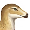
In Figure I you can see the skull of a red dragon in lateral view. Note how the skull is lightened by the large naris (Na.), antorbital fenestra (Ant.f), postorbital fenestra (Post.f.) and the mandibular fenestra (Man.f). The eqully large orbit (Orb.) houses the sclerotic ring (Scl.r) that supports the eyeball. Eudraconids have interlocking paracanines, one in the premaxilla (Prem.), one in the maxilla, and their counterparts in the dentary (Den.). The cornual process (Cor.pr), formed from a fusion of cranial bones forms the base for the laterally flattened ceratinous horns. These and other ceratinous growths and muscular anatomy are shown in black.
Figure II shows you the Pectoral Girdle and trunk of an eudraconid dragon (ribs and alar humerus not depicted). Note how the scapulacoracoid, formed from a fusion of posterior scapula (Post.sc) and coracoid (Post.c.), attaches to the notarium (Not.) formed from fused dorsal vertebrae and the enlargened sternum (St.) or breastbone. Together they keep the rib cage rigid and prevent its compression during flight. Also attached to the scapulacoracoid near the glenoid fossa (Gl.fs) is the anterior scapula (Ant.sc.). The humerus (Hum.) and coracoid (Ant.c) of the anterior limb pair, as well as the pubic bone or ilium (Ill.[sic]) are also partly depicted here.
(This pic is up in my Elfwood gallery with the same description as well, but I wanted to put a higher res version here with the rest of my Aren stuff for easy access.)
Image size
600x762px 133.76 KB
© 2011 - 2024 Osmatar
Comments12
Join the community to add your comment. Already a deviant? Log In
Thank you for giving the wings and front legs seperate sockets.